What is qPCR testing?
Used to quickly identify pathogens and antimicrobial resistance markers in a small sample, qPCR is indispensable for the modern point-of-care molecular diagnostics.
Polymerase Chain Reaction (PCR)
Polymerase Chain Reaction (PCR) is a molecular diagnostic method used to increase the amount of DNA or RNA in a sample. This is necessary in molecular diagnostics because there isn’t enough bacterial and viral DNA or RNA in a typical clinical sample for detection, even in a laboratory setting.
While there are other methods of nucleic acid amplification, PCR is routinely used to test the human population for COVID-19 and other pathogens. In veterinary medicine, PCR can be used to identify bacteria that cause infectious conditions like urinary tract infections.

How Does PCR Work?
PCR testing, or Veterinary PCR testing, works by mixing a clinical sample that may contain a pathogen of interest with a customized biological test mixture or ‘assay’. The assay mixture is then placed into a thermal cycling instrument that, by varying the temperature over a prescribed time, causes the DNA to be multiplied into millions of copies in about an hour.
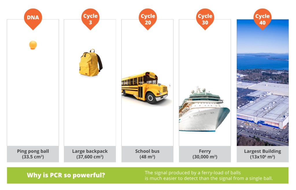
A simple way to imagine this is by using ping pong balls as DNA molecules. If the starting DNA was a ping pong ball with a unit volume of 33.5 cm3 and the equivalent volume was amplified by PCR, then at cycle 3 the amplified volume would fit in a large backpack (37,600 cm3). Continuing this cycle will fill a yellow school bus with 48 m3 of ping pong balls at cycle 20, a ferry boat with a volume of 30,000 m3 at cycle 30, and by the time the PCR reaction is complete at cycle 40, there would be an equivalent volume of 13 x 106 m3 of ping pong balls to fill the largest building in the world (Figure 1).
A signal produced by a ferry-load of DNA molecules is much easier to detect than from a single DNA molecule, which is why PCR is so essential in molecular diagnostics.

Real-time PCR (qPCR)
qPCR refers to real-time PCR or quantitative PCR, which allows for simultaneous amplification and detection of DNA in real-time. This supports faster detection of target DNA sequences, increased testing sensitivity, and lower risk of cross contamination, all factors driving the increasing adoption of qPCR to rapidly diagnose clinical conditions in both human and animal populations.
How Does qPCR Work?
qPCR works by amplifying target DNA to levels in which a detectable signal can be observed. The reaction is prepared by mixing a small amount of a sample (e.g., saliva or urine) with the building blocks needed to make copies of the DNA sequence.
What sets qPCR apart from older methods is that the reaction mixture also contains fluorescent probes, or DNA binding dyes. If the sample contains the target DNA sequence (e.g., DNA from a pathogen such as Staphylococcus), the binding dyes will attach, activate, and produce a detectable signal.
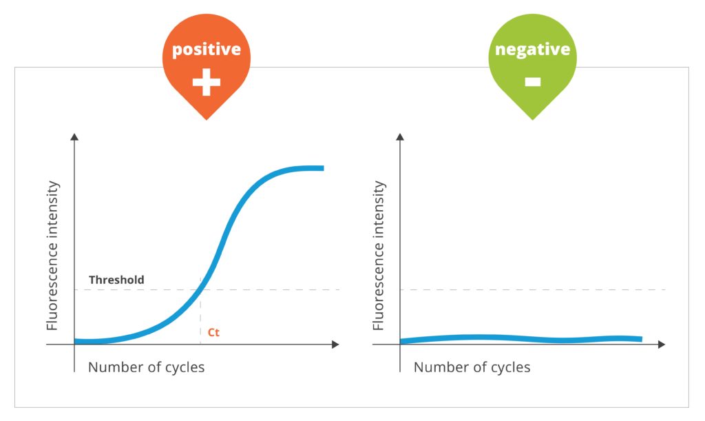
The photocopied or ‘amplified’ DNA is monitored by measuring light from probes specific to the target DNA of interest through a process called fluorescence. The results determine the presence or absence of a molecular target (e.g., pathogen). In the case of a sample with an abundance of a virus, the measured signal would increase rapidly over time. However, if there is no or a very low concentration of DNA in the original patient sample, no signal would be observed, indicating a negative result (Figure 2).

Steps in the qPCR Process
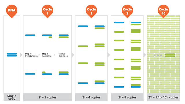
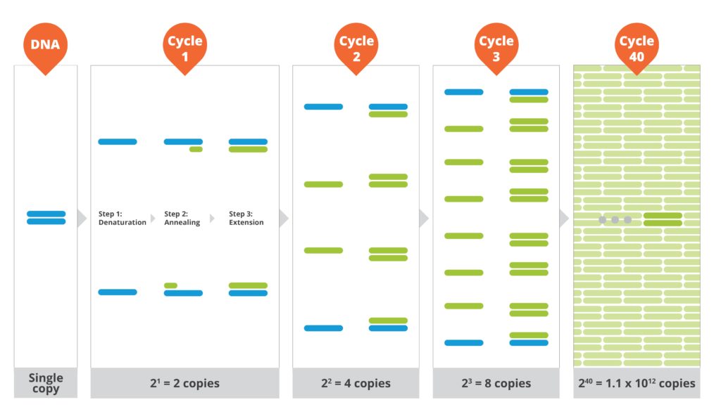
The qPCR process consists of three steps and requires controlled temperature cycles that are repeated approximately forty times.
- The first step is the denaturation stage, which occurs at an elevated temperature to enable separation of the double-stranded DNA.
- Next is the annealing stage, which occurs at a lower temperature allowing the primers to bind to the flanking regions of the target DNA and create the photocopied or amplified copy.
- The final stage is the extension phase, which is performed at an elevated temperature and a new strand of DNA is made.
The DNA amplification process continues over approximately forty thermal cycles and until all reaction components are consumed (Figure 3).

What Information Does the qPCR Amplification Curve Provide?
An amplification curve is generated from the real-time detection of the qPCR cycle and contains the initiation, exponential, and plateau phase as they relate to the cycle number and fluorescence emitted. A typical amplification curve is shown in Figure 2. Two values are particularly useful in a qPCR curve.
- The first value is the threshold line, which is where the fluorescence reaches a level higher than the baseline.
- The second value is the Ct value and the point at which the reaction curve crosses the threshold. The Ct value indicates the number of cycles required to detect a positive signal.
A positive result is shown as an increase in the amplification curve. A negative result is shown as a flat line and obtained in the absence of target DNA in the sample.
Applying qPCR techniques in molecular diagnostics provides a powerful tool for the detection of a wide range of pathogens in human or veterinarian patients.
